HAEMODIALYSIS ACCESS MAINTENANCE
Patient Education Material
Haemodialysis is a method used to remove fluids and waste from the blood in patients with kidney failure. This requires that the patient’s blood flows into the haemodialysis filtering system. There are three main access routes for this: intravenous catheters (thin tubes which are placed in a blood vessel); arteriovenous (AV) fistulas (connections created between an artery and a vein); and synthetic grafts (artificial veins). Intravenous catheters are only used for short periods.
The most common access method is an AV fistula. This is a channel that is created by surgically joining an artery and a vein. Synthetic AV grafts are artificial vessels connecting an artery to a vein and tend to be used when the patient is unsuitable for an AV fistula.
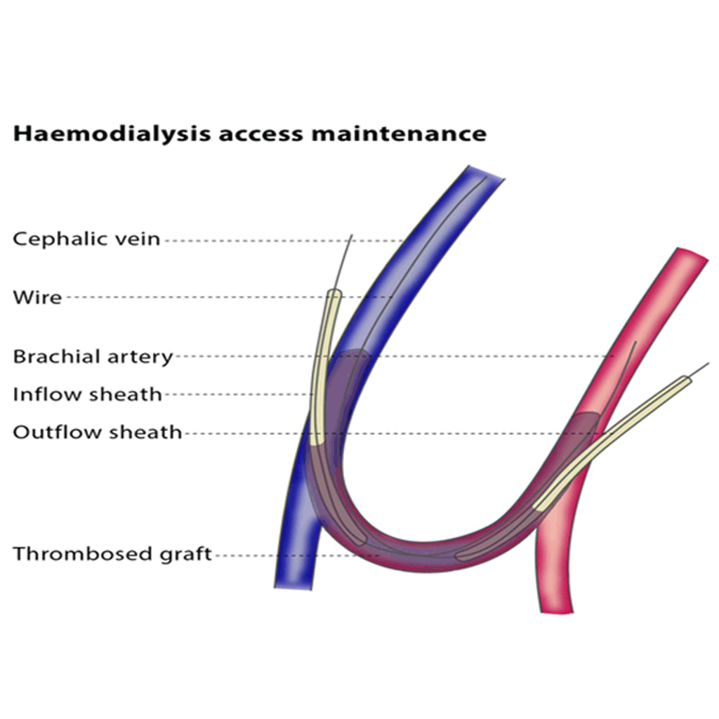
Both AV fistulas and synthetic grafts may become narrowed, thrombosed (obstructed by blood clots) or blocked. In order to keep them clear, you may undergo a percutaneous (through your skin) treatment, such as percutaneous thrombolysis, thrombectomy, balloon angioplasty and stenting. The process of keeping the AV fistula or graft clear is called haemodialysis access maintenance.
Haemodialysis access maintenance procedures are performed on an outpatient basis and use fluoroscopic guidance. Your vital signs will be monitored during the procedure.
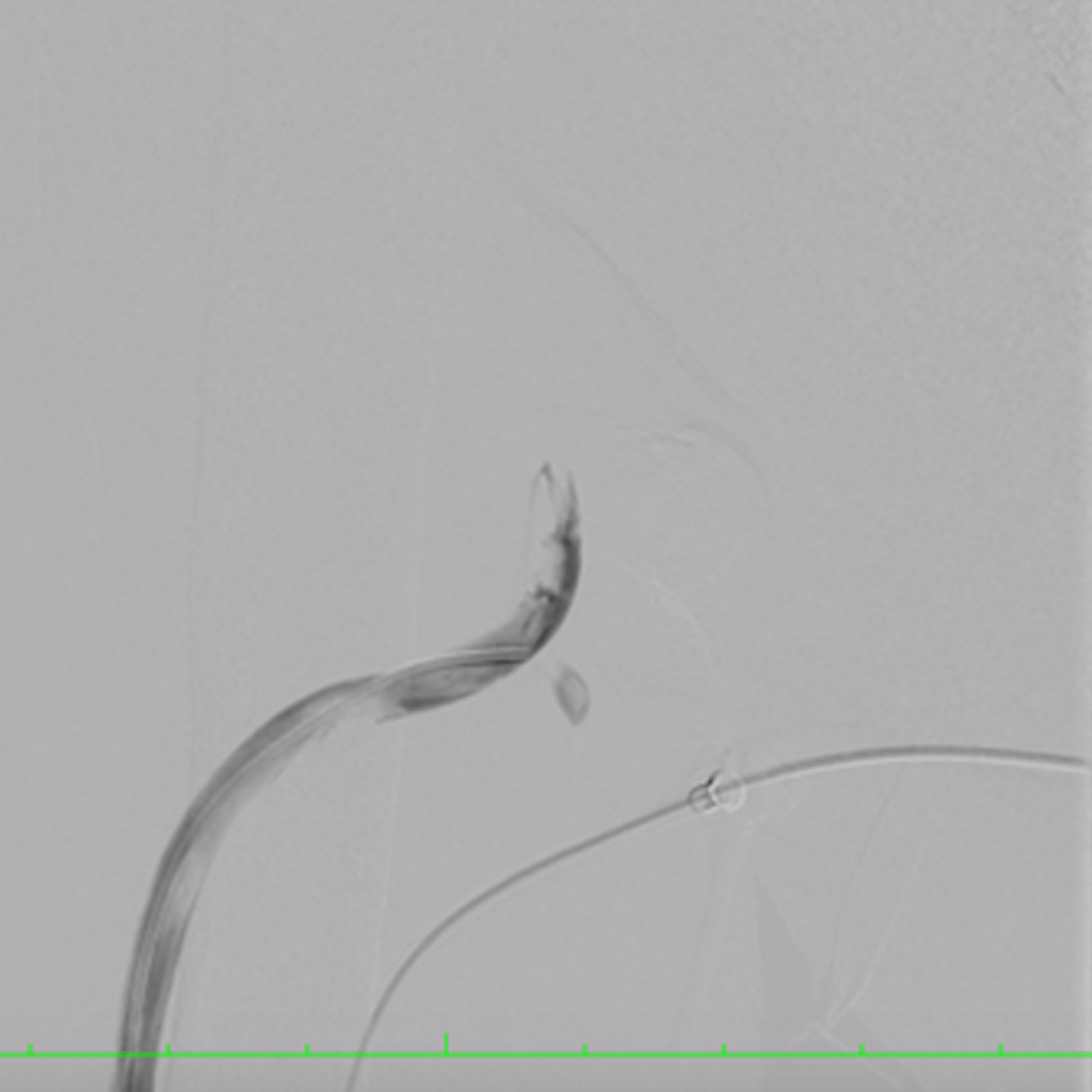
You will undergo local anaesthesia. The interventional radiologist will cut a small incision in your skin, and will then insert a catheter and a guidewire into your AV fistula or synthetic graft. If there is a blood clot present, the interventional radiologist can remove it by performing a procedure called a thrombectomy.
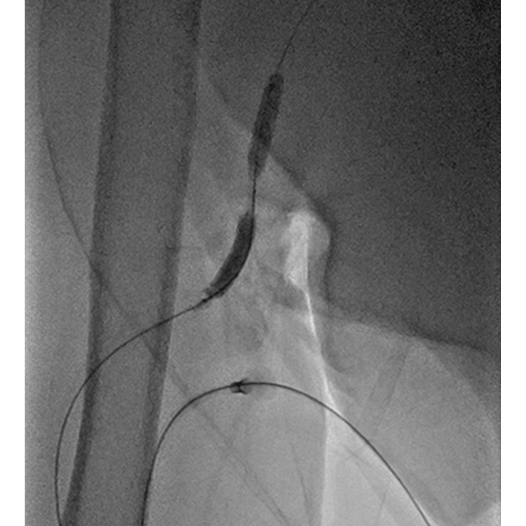
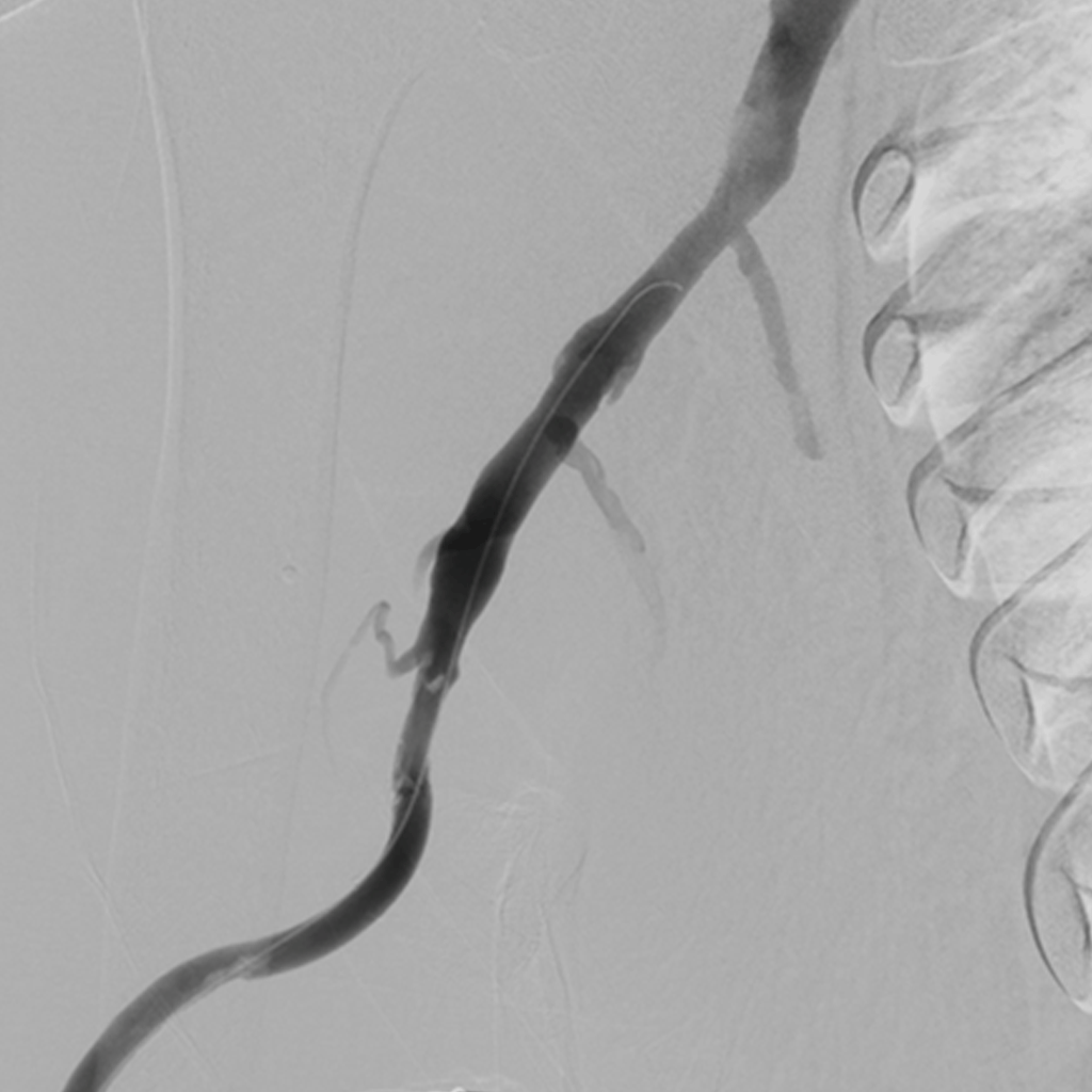
In order to accurately diagnose the location and severity of any narrowing in the AV fistula, the interventional radiologist will inject a contrast material (dye) into the fistula, the inflow artery and the outflow vein, and will perform an angiography on the area. The interventional radiologist will then insert a tiny inflatable balloon into the narrowed area using a guidewire. As the balloon expands, it gently widens the vessel wall and restores the vessel’s diameter. In some cases, the interventional radiologist will place a stent (a specially designed metal tube) in the vessel to support the vessel walls and keep them open.


If there is a blockage or narrowing in an AV fistula or synthetic graft, it will not be possible for blood to flow to the haemodialysis machine. The development of percutaneous maintenance techniques has prolonged the life of AV fistulas and grafts. As a result, haemodialysis access maintenance procedures reduce the need for temporary central catheters and help to preserve the vessels in the kidney.
Kindly contact:
- One PKLI Avenue, DHA, Phase-6, Lahore, Pakistan.
- info@pkli.org.pk
- +92 42 111 117 554

